
Neuroanatomy II: Understanding the Ventricles
In our previous article, Learning the Brain: Neuroanatomy Basics for Understanding Hydrocephalus, we discussed the anatomy of the brain and what happens to the brain in hydrocephalus. But one thing that is important to know is where all of this is happening and why. Oftentimes, you’ll hear your ventricles being referred to as “lateral”, “third”, “fourth”, or even “aqueduct”. This can be confusing when you’re trying to figure out where in your brain these different areas are, and how it relates to hydrocephalus treatment. The goal of this article is to highlight the difference between each ventricle, their locations, and their importance to hydrocephalus.
The Ventricles and the Production of CSF
Inside your brain, you have a series of ventricles, or fluid-filled cavities. These ventricles are filled with cerebrospinal fluid (CSF) that is constantly moving and working to cushion the brain and remove toxins. The CSF flows in one direction, creating a type of current that surrounds your brain and spinal cord. Because the CSF flows in one direction, this means that if there is a blockage or interruption, it creates a buildup like a traffic jam. It is important to note that this traffic jam can occur in different locations in your ventricles depending on the cause. This buildup of CSF is known as hydrocephalus.
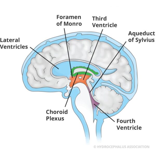
When you’re learning about hydrocephalus and treatment options, you may hear someone talk about the different ventricles and nearby structures. These structures together are known as the ventricular system. If we look at the journey of CSF through the body as a commute on a road, it starts in the lateral ventricles (a connected pair with one half on each side of the brain) and includes the choroid plexus. The choroid plexus is a small piece of tissue that floats in the lateral ventricles. It has many roles in the ecosystem of the brain, but its primary function is the production of CSF.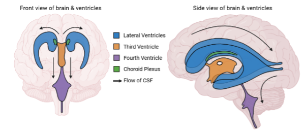
After being produced in the lateral ventricles, the CSF flows around from the middle of the brain to the bottom, and through the third ventricle and lastly fourth ventricle. The third ventricle is a narrow space that sits along the midline, or the invisible line that separates the brain into two halves. The third ventricle acts as a meeting point for the CSF coming from both the lateral ventricles. CSF flows out of the third ventricle through the aqueduct to the fourth ventricle.
The fourth ventricle, downstream of the third, sits right above the spinal cord and is where the CSF exits the ventricular system to flow around the outside of the brain or around the spinal cord to eventually be absorbed.
Treating Hydrocephalus in the Ventricles
There are many different types of hydrocephalus, and each patient’s situation is different. With this in mind, there are two primary treatments that involve intervention within the ventricles: shunts and endoscopic third ventriculostomies (ETVs).
Watch the video below to learn more about how hydrocephalus is treated:
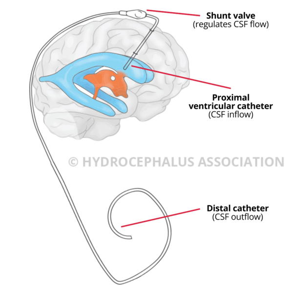
To recap the video, shunt systems work by placing a small, flexible tube called a catheter into one of the lateral ventricles to drain excess CSF to another part of the body. The fluid is often sent to the abdomen or heart because the body can absorb it there safely.
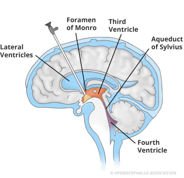
Alternatively, in addition to or instead of a shunt, an endoscopic third ventriculostomy (ETV) is performed. An ETV is a procedure in which the surgeon creates a small hole in the third ventricle to bypass a buildup or blockage and increase the flow of CSF out of the ventricular system. This is done with an endoscope, or a camera on the end of a long, flexible tube.
Additionally, some patients that are eligible to receive an ETV may also have a choroid plexus cauterization (CPC) performed. As mentioned above, the choroid plexus is located in both lateral ventricles, where its primary job is to constantly produce CSF. Due to its productive nature, some surgeons find thatsmall parts of the choroid plexus (called cauterization or coagulation), alongside the ETV can be an effective treatment for certain patients. CPC can help by reducing the amount of CSF the brain makes, aiming to prevent fluid from building up too quickly.
While every person with hydrocephalus may have differences in severity and ventricle size, the overall structure and location of the brain’s ventricles is the same for most people. To recap, the two lateral ventricles are on each side of the brain, and they both flow into a single ventricle called the third ventricle. The CSF that ends up in the third ventricle will continue flowing down towards the spinal cord through the aqueduct and fourth ventricle, and from there will either travel up and around the outside of the brain, or down and around the spinal cord.
This video from 2-Minute Neuroscience provides a brief summary of what we have covered in this article:
Why This Matters
Understanding how CSF moves through the ventricles helps explain how hydrocephalus and its treatments work. When this flow is blocked, or fluid builds up faster than it can be absorbed, pressure can increase and symptoms can develop. Treatments like shunts and ETVs are designed to restore balance by redirecting fluid or creating a new pathway for it to flow. This overview of the ventricular system is based off our current understanding of CSF dynamics, but our understanding of CSF flow grows as ongoing research in this area of medicine continues.
If you’d like to explore this topic further, check out our related article: Learning the Brain: Neuroanatomy Basics for Understanding Hydrocephalus