
Get to Know Dr. Engin Deniz: HA 2019 Innovator Award Grantee
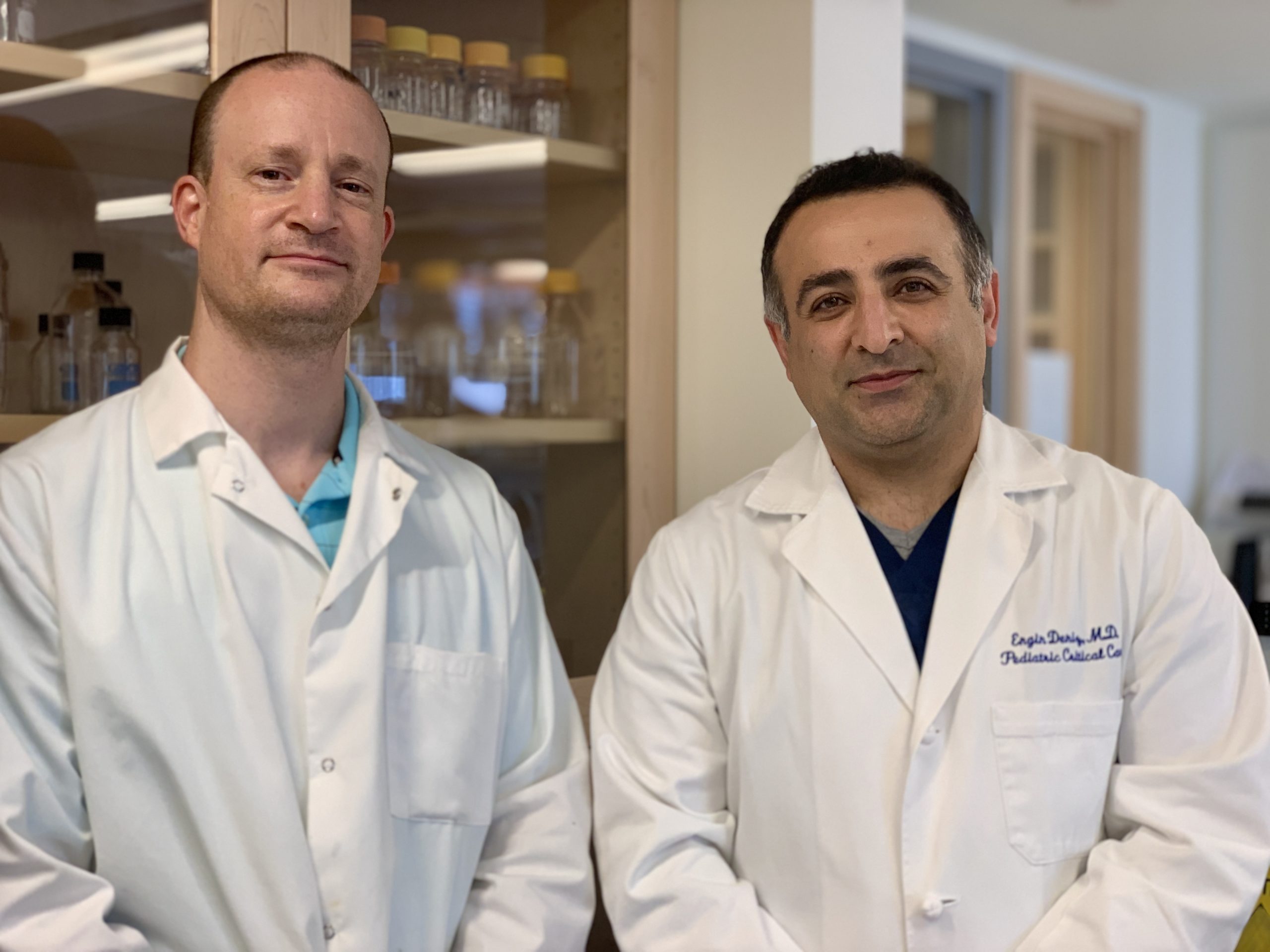
Dr. Deniz (pictured right) with research assistant, Stephen Viviano.
In January, we announced our 2019 Innovator Award grantees. The Innovator Award is designed to provide seed funding for bold and innovative work with the potential to transform hydrocephalus research. In the final installment of our Meet the Innovator Award Grantees Blog Series, we interviewed Dr. Engin Deniz, one of four scientists who received a 2019 Innovator Award. Dr. Deniz is an Assistant Professor in the Division of Critical Care Medicine at Yale University. Through his research, he hopes to determine how cilia, small hair like structures that move cerebrospinal fluid (CSF), contribute to post-traumatic hydrocephalus.
What sparked your interest in hydrocephalus research?
I’m a pediatrician and a critical care physician and I’m interested in birth defects. We’ve been studying a wide range of birth defects and congenital hydrocephalus was one of them. The frog model that we’ve been using to study congenital heart disease or other birth defects turned out to be a powerful model to study hydrocephalus. My colleague Dr. Kristopher Kahle, one of the pediatric neurosurgeons here at Yale, has been studying genetic causes of hydrocephalus and has found novel genes that have never been implicated in hydrocephalus. We now have plenty of genes that require functional screening before they are studied in-depth. So I built the frog model as a useful tool to functionally screen these genes and look in detail at the pathogenesis of hydrocephalus and that’s when my interest first took off about three years ago. So I’ll be using the frog system as the focus in my biology work.
What do you find most interesting about hydrocephalus research?
The children that I’m taking care of motivate me the most. We see a spectrum of patients presenting with hydrocephalus. The most common are patients with post-infectious hydrocephalus, but we also see many babies born with hydrocephalus. The impact of hydrocephalus on their lives is immense. My second motivation is to see if we can come up with a useful medical treatment, so we don’t have to rely on the old fashion surgical options that we have now. The treatment that we have for hydrocephalus has been failing. There are only two surgical options, and the main procedure – inserting a shunt to drain fluid – was developed 50 years ago. Unfortunately, many of our patients come to the ICU with reoccurring infections and shunt malfunctions, and they can go through multiple revisions. Besides surgical options, we don’t have much to offer these families. So what is the genetic basis, and how can we help these families in terms of prognosis? We aim to identify specific developmental pathways and have more insight into how we can start thinking about reversing this or simply preventing it by finding a potential molecular underlying problem that we can target.
How long have you been researching hydrocephalus?
Nearly 3 years but I have been involved in biology for 10 years now.
What do you hope to learn from your research funded by the Innovator Award Grant? What questions do you hope your research will answer?
The Innovator Award has been fundamental in what I’m doing. It has given me two perspectives. First, these type of awards motivate you. The second part is that I’m focused on a particular type of hydrocephalus that is related to ciliopathies. Multiple questions remain unanswered in this field. For example, what is the role of cilia in CSF dynamics during the early embryonic stages? How is the circulation of CSF compromised in patients born with hydrocephalus? How is this effected following intracranial bleed? The frog model system gives us a little bit of advantage in studying this very early embryonic CSF physiology. We can access the whole ventricular system in the frog system at the early stages of ventricle formation. So, my focus is to understand what the role of cilia is in developing hydrocephalus. We’re looking at the genetics of cilia and the physiology of cilia driven CSF circulation, which can be tracked in the frog system. What I’m excited about is the fact that we can look at the relationship of the pathogenesis of hydrocephalus in ciliopathies in the most detailed fashion.
What makes your project unique?
Pairing Optical Coherence Tomography imaging with the frog system to study hydrocephalus is entirely novel. We have a different perspective where we can assess embryonic CNS development and CSF dynamics in real-time. The ease of accessing the early embryonic brain gives us much fundamental insight. For example, how aqueductal stenosis forms or how CSF circulation evolves – you can watch that in real-time in this model. That’s the unique position we’re in, and we’d like to capitalize on that moving forward. The idea is to find potential genetic causes that would lead to hydrocephalus, and using the frog model system we aim to identify the earliest time point during development that deviates from normal to better understand the disease’s mechanism.
How important is HA’s Innovator Award grant for your project?
The innovator award was motivating and led us to an NIH R21 award. The grant allowed us to further focus on publishing our paper to layout the frog model system’s groundwork to study hydrocephalus. We’ve made significant progress, and that’s promising.
What is the long-term plan for the project?
The long-term plan is to focus on the role of the ependymal cilia in hydrocephalus. There is a plethora of cilia in the brain. Some of them are destined to beat and contribute to CSF circulation, and there is a second group of cilia that are immotile and mainly act as sensory organelles. The relationship between these cilia is crucial for brain development, yet our understanding of how is incomplete. The long-term idea is to decipher the relationship between these two types of cilia in early embryonic brain development and determine how ciliary defects lead to hydrocephalus. We believe the genetically tractable frog model system is ideal for this purpose. The frog model is rapid and cost-defective, ideal for functional screening of the novel genes derived from human studies. Most importantly, we can visualize the cilia driven embryonic CSF circulation of the entire tadpole ventricular system in a non-destructive fashion, enabling us to study the interwoven relationship between neurogenesis and the complex early CSF circulation.