
Medical Device Development and Evolution: A Comparative Study
 By Marvin Sussman, PhD
This article with photos and graphs can also be found in the latest edition of our print newsletter, Pathways.
The first commercially-available shunt was introduced almost 60 years ago. Over the past 60 years, significant developments and evolution of medical device technology has occurred in many fields of medicine. This article compares and contrasts the evolutionary changes that have occurred in the field of hydrocephalus with the revolutionary changes that have occurred in two other fields: cardiac pacing and radiation oncology. What we find is that while the shunt has seen modifications that have had positive impacts for patients, more is needed if we are going to move the management and, one day, the cure of hydrocephalus into the 21st century.
A Brief History of Three Medical Devices: Cardiac Pacing, Radiology, and the Shunt
Cardiac Pacing
The first cardiac pacemaker was an external stimulator designed and built-in 1950 based upon design input by a cardiothoracic surgeon. A large external device, the size of a cart, was built using vacuum tube technology; it provided transcutaneous pacing using electrodes on the skin. It was crude and painful to use and, being powered from an AC wall socket, had a potential hazard of electrocution by inducing an abnormal heart rhythm. The engineers began to tackle the size of the unit as well as the power source.
The first commercially available silicon transistor was developed in 1956. This led to the miniaturization and rapid development of practical cardiac pacemakers. In 1958, the first wearable external, transistorized pacemaker was used for a patient. It was housed in a small plastic box with controls for rate and output voltage adjustment. It was connected to wire leads which passed through the patient’s skin to electrodes attached to the heart surface. The first fully implantable pacemaker into a patient occurred later that year, with electrodes attached to the heart’s myocardium. The device lasted for three hours; a second device was implanted and lasted for two days. In 1959, the successful use of a temporary transvenous pacer was first demonstrated. In 1960, primary cell mercury batteries were introduced that could power an implant for up to 18 months. These implantable devices all suffered from poor reliability and short battery life associated with mercury batteries.
In the late 1960s, several companies developed isotope-powered pacemakers, but they were surpassed, in 1971, by lithium-iodide cell battery which more than doubled pacer battery life and provided advance warning of impending failure. This was coupled with the introduction of heretically sealing technology to overcome the problem of the intrusion of body fluids into the unit, which was also affecting the reliability of the device.
More recent advances in the implanted pacemaker have focused on the transition to microprocessor-controlled pacemakers. This led to pacemakers that improved the heart’s pumping efficiency, then eventually evolved into the ability to adjust the base pacing rate using rate response algorithms. Pacemakers now provide information via wireless transmission which the clinician uses to adjust the pacemaker function to the patient’s specific requirements and provides data for inclusion in the patient’s record.
Radiation Oncology
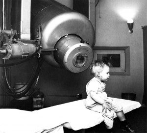
Radiation oncology, as a field, was born not long after the discovery of x-rays in 1895. However, a new era of therapy occurred in 1956, when a linear accelerator, a device used by radiation physicists for research purposes, was adapted for use as a tool for fighting cancer. A 2-year-old boy suffering from retinoblastoma, a cancerous tumor in his eye, was the first patient to be treated using X-rays produced by a research linear accelerator modified by physicists for medical use. This treatment saved the child’s sight; for the rest of his life his vision remained intact. In 1958, the first commercial medical-use linear accelerator (“Linac”) was introduced.
By the 1960s, high-energy (megavoltage) Linac treatment machines heralded the next generation of treatment technology. They were capable of producing high energy, deeply penetrating radiation beams, which allowed the treatment of tumors deep inside the body without excessive damage to the overlying skin and other normal tissues. In subsequent years, radiation oncology experienced multiple technologic revolutions. By the late 1980s, computer tomography (CT) was linked with Linacs to create CT-based treatment planning. Software algorithms produced sophisticated treatment plans for radiation therapy allowing three-dimensional conformal radiotherapy (3D-CRT). This planning reduced radiation doses delivered to surrounding normal tissues, greatly improving the quality and delivery of radiotherapy. However, stray radiation scatter to surrounding healthy tissues remained a problem.
In 1992, Intensity Modulated Radiation Therapy or IMRT was introduced. Perhaps the most important “revolution” in radiation oncology to date, IMRT conforms the prescription radiation’s therapeutic dose to the volume shape of the defined target in 3 dimensions, thereby reducing the volume of normal tissues receiving high radiation doses. This reduces the potential side effects of radiation treatment. More importantly, IMRT also provides a method to safely escalate the radiation dose, allowing re-treatment of previously irradiated patients, potentially improving cure rates and giving repeat-treatment-patients the option of additional radiation treatments. IMRT has become standard practice for a wide range of tumors.
In 1998, ultrasound was combined with treatment planning so that radiation can be delivered to targets that move daily, such as the prostate. Currently, the field is undergoing another technologic revolution with the development of Image-Guided Radiation Therapy (IGRT) which provides the ability to perform cone-beam CT scans of the patient while the patient is on the treatment table allowing treatment to be adapted to tumor shape changes occurring over multiple radiation treatment sessions. This is expected to translate into higher cure rates and lower rates of toxicity.
Hydrocephalus Shunts
The development of the biocompatible silicone elastomer – a type of plastic that could be implanted in the human body – allowed the introduction of hydrocephalus shunts. The first clinically-viable, valve-regulated hydrocephalus shunt, introduced in 1952, ushered in the modern era of hydrocephalus surgery and treatment. At that time, there was no effective medical intervention for hydrocephalus. In that year, two neurosurgeons working in conjunction with a hydraulics technician whose child had hydrocephalus, successfully implanted a silicone elastomer, ventriculojugular shunt regulated by a ball-in-spring valve design inserted at either end of a flexible piece of tubing. This device regulated and released controlled amounts of the cerebrospinal fluid (CSF) from the brain into the heart’s right atrium.
At about the same time, another neurosurgeon produced a one-way slit valve made of silicone elastomer. The development of the valve system combined with the application of new biocompatible materials allowed for the safe and reliable diversion of CSF without many of the complications of unregulated (systems without valves) CSF drainage. Other neurosurgeons soon introduced the concept of ventriculoperitoneal procedures, the distal end of the tubing ending in the abdominal cavity, in which these newer devices were used.
While the early shunts allowed for the management of hydrocephalus, patients still faced numerous repeat surgeries due to shunt malfunction and/or shunt infection. Focus turned to the creation of a more sophisticated valve mechanism that would control the rate of CSF flow through the shunt, taking into account the variables caused by regular daily activity.
The 1970s saw the introduction of valves that addressed overdrainage or siphoning of CSF caused by an individual’s postural (standing, sitting, or lying down) change and vasogenic (blood flow) influences. As an example, moving to a standing posture causes a siphon or sucking effect, essentially “pulling” fluid out of the brain or lumbar region. The anti-siphon device was introduced in the early 1970s, followed by the gravity compensating mechanism in 1976. Flow regulated shunts were introduced in 1987. These valves maintain the drainage flow rate close to the rate of CSF secretion, regardless of patient position and other conditions that normally promote overdrainage.
The problem remained that when these devices were not draining fluid properly, the individual had to undergo brain surgery to adjust or replace the shunt. The mid-1980s heralded in the programmable shunt. These shunts can be adjusted noninvasively using a magnet. The first magnetically-adjustable valve was marketed in 1984.
Advances also addressed reducing infection rates both during and after surgery. In 1976, the first commercially-sterilized shunt came to market. Antibacterial catheter coatings became commercially available in 2001. These advances in shunt construction have been coupled with standardized procedures produced by the Hydrocephalus Clinical Research Network (HCRN) that have dramatically reduced infection rates in the pediatric population.
There are now literally hundreds of options for valves, proximal and distal catheters. Further, device modifications to minimize overdrainage, antibacterial coatings, and magnetically-adjustable valves for fine-tuning CSF flow rates, are all evolutionary modifications of the original clinically-viable shunt design.
Progress in imaging technology has allowed clinicians to treat hydrocephalus with greater success and safety. In the 1980s endoscopes again found a role in neurosurgery. The benefits include more accurate placement of ventricular catheters and creation of third ventriculostomies, primarily for aqueductal stenosis, in carefully-screened patients. Stereotactic localization, a minimally invasive surgical technique, led to more functional and safer approaches for the CSF drainage.
Comparing Technology Development Over the Sixty Year Period
Great advances and achievements have been made over the course of the past six decades in many medical fields. Regrettably, the progress and advancements in the field of hydrocephalus has been slow and has not kept pace with those that have been achieved in other fields.
Cardiac pacemakers evolved from a cart-sized, AC line-powered device that was only adjustable for output voltage and rate to a fully-implantable, coin-sized device with long-life batteries, sensing circuitry and non-invasive adjustability for parameters such as rate, pulse width, output current, parameters sensed, telemetry, etc.
Radiation oncology has progressed from providing treatment with a re-tuned research linear accelerator to one with sophisticated radiation treatment planning software that allows the delivery of radiation with pinpoint accuracy, combining radiation delivery with virtually real-time image guidance allowing radiation to be focused on a tumor whose volume is changing based upon the treatment delivered.
Hydrocephalus management moved at a much slower pace. Shunts have changed relatively little compared to the advancements made in other fields. Shunt tubing is made from the same material, silicone elastomer, although it has been made radiopaque to facilitate visualization on X-ray. Valves work based upon the same differential pressure principles across the valve and valve adjustments are by-and-large in discrete adjustment steps for setting the valve resistance when adjustable valves are used. Valves are available in multiple pressure ranges (as was the case with the first shunts). Magnetically-adjustable valves have taken over the field but, once set, they function in the same way that the first shunt functioned. Some shunts incorporate antibacterial coatings which appear to minimize infection during the immediate post-operative period. After this period, the risks of infection are similar to untreated shunts.
The shunt system remains relatively unchanged consisting of an inflow (proximal) catheter, a regulating device (valve) and an outflow (distal) catheter. No commercially available shunt contains a sensor nor is there closed-loop, feedback control mechanism to adjust shunt function to the changing patient’s needs. Telemetry of information that is now routinely available in cardiac pacemakers or neural stimulators is unavailable in shunts. There are no active shunts, although implantable pumps have been available for several decades.
Conclusion
Clinicians in the 21st century will be required to continue to face the challenges presented by hydrocephalus. Treatment, until now, has focused on the management and alleviation of symptoms rather than the arrest and cure of the underlying processes that cause the imbalance between CSF production and absorption that cause the hydrocephalus condition. Treatment is plagued with one of the highest complication rates of any medical implant. Routine complications of the treatment modality (the shunt) are infection, obstruction, and overdrainage to name a few. Although some (regrettably the minority) of the patients with shunts can go for years without complications, even those lucky few are potentially one shunt malfunction away from a major crisis. At any time, and without warning, a shunt complication can require emergency intervention which may turn into a course in which many shunt surgeries are necessary.
Medical device manufacturers are commercial enterprises responsible to their shareholders to produce a profit. Product-development decisions are based upon market size and return-on-investment calculations.
The integration of the discoveries in basic science and clinical innovation from other fields will, hopefully, lead to the revolutionary technological and therapeutic developments that have brought fields such as cardiac pacing and radiation oncology into the 21st century. Due to the hydrocephalus market’s comparatively small size against those of the cardiology or radiation oncology markets, revolutionary advances coupled with significant investments may not be commercially justifiable in and of themselves. Advancements in research in other fields, such as gene therapy, molecular biology, and neural regeneration, may be applicable to hydrocephalus and, thus, assist in moving development forward. Technological developments from other fields, such as closed-loop feedback sensing technologies developed for cardiac pacing and implanted neural stimulation devices and implantable pumps for intrathecal and other drug delivery, may be applicable to the management of hydrocephalus to eventually produce a “smart shunt.” The concept of a functional cure rather than palliation may become achievable.
The Hydrocephalus Association has undertaken the research initiative to raise funds to attract and support the best and the brightest to develop revolutionary solutions to the problems that have plagued those impacted by hydrocephalus.
 Marvin L. Sussman, Ph.D. is an expert in the field of medical device development and regulation with over 35 years of experience in the Medical Device Industry. He currently consults for a number of companies in medical device and market development, regulatory affairs, and quality assurance. He has participated in the defense of medical product litigation related to hydrocephalus shunts and other medical devices. Prior to becoming a consultant, he held a number of senior executive positions at NOMOS Corporation, where he guided the development of the organizational structure, supervised the implementation of their quality system through ISO 9001 certification, and developed the strategy that gained regulatory clearance for the first devices in the field of Intensity Modulated Radiation Therapy (IMRT). Before joining NOMOS, he held a number of senior management positions at CORDIS Corporation where he supervised the development of a number of neurosurgical products (hydrocephalus shunts, neural stimulators, CSF drainage devices, etc.) through the 510(k) or PMA process and foreign regulatory approvals and market introduction. Dr. Sussman served as the co-chair of the AAMI Neurosurgery Committee, the AAMI ICP, and ASTM CNS Shunt Standards Committees and was the industry representative on the FDA Neurological Devices Panel twice. Dr. Sussman holds a B.S. in Biology from Brooklyn College and an M. S. in Physiology from Long Island University. His doctoral degree and postdoctoral studies were from the University of Miami Miller School of Medicine in Neurophysiology and Biophysics. He holds a number of patents for shunt and other devices in Neurosurgery and the sterile packaging/testing of medical devices. Dr. Sussman has been a member of the Board of Directors for 13 years, having advised on numerous internal projects that have moved the HA agenda forward. Dr. Sussman has shared his wealth of knowledge regarding hydrocephalus and medical devices to our membership in the form of papers, articles, blogs, and presentations. Marvin L. Sussman, Ph.D. is an expert in the field of medical device development and regulation with over 35 years of experience in the Medical Device Industry. He currently consults for a number of companies in medical device and market development, regulatory affairs, and quality assurance. He has participated in the defense of medical product litigation related to hydrocephalus shunts and other medical devices. Prior to becoming a consultant, he held a number of senior executive positions at NOMOS Corporation, where he guided the development of the organizational structure, supervised the implementation of their quality system through ISO 9001 certification, and developed the strategy that gained regulatory clearance for the first devices in the field of Intensity Modulated Radiation Therapy (IMRT). Before joining NOMOS, he held a number of senior management positions at CORDIS Corporation where he supervised the development of a number of neurosurgical products (hydrocephalus shunts, neural stimulators, CSF drainage devices, etc.) through the 510(k) or PMA process and foreign regulatory approvals and market introduction. Dr. Sussman served as the co-chair of the AAMI Neurosurgery Committee, the AAMI ICP, and ASTM CNS Shunt Standards Committees and was the industry representative on the FDA Neurological Devices Panel twice. Dr. Sussman holds a B.S. in Biology from Brooklyn College and an M. S. in Physiology from Long Island University. His doctoral degree and postdoctoral studies were from the University of Miami Miller School of Medicine in Neurophysiology and Biophysics. He holds a number of patents for shunt and other devices in Neurosurgery and the sterile packaging/testing of medical devices. Dr. Sussman has been a member of the Board of Directors for 13 years, having advised on numerous internal projects that have moved the HA agenda forward. Dr. Sussman has shared his wealth of knowledge regarding hydrocephalus and medical devices to our membership in the form of papers, articles, blogs, and presentations. |