
Understanding External Ventricular Drains (EVDs) in Hydrocephalus Management
External Ventricular Drains (EVDs) are important medical devices where a ventricular catheter is inserted directly in the brain and redirects (drains) cerebrospinal fluid (CSF) to an external collection system. It is frequently used in head injury (as the system can also monitor the pressure) and while treating infections, bleeding, or other complex shunt issues. This article explains how EVDs are used specifically for hydrocephalus, when they are necessary, and what to expect during their use in the hospital.
What is an External Ventricular Drain (EVD)?
An EVD is a thin, flexible tube that is inserted into the ventricle (fluid-filled space) in the brain to drain CSF. In hydrocephalus, where the flow of CSF is blocked or disrupted, this fluid can build up and cause dangerous pressure inside the skull. The EVD system can:
- Relieve pressure by draining the CSF outside of the body.
- Measure the pressure in the brain (intracranial pressure or ICP).
- Collect CSF samples to check for infections or other issues.
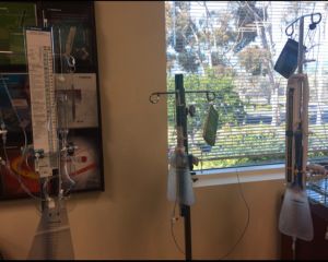
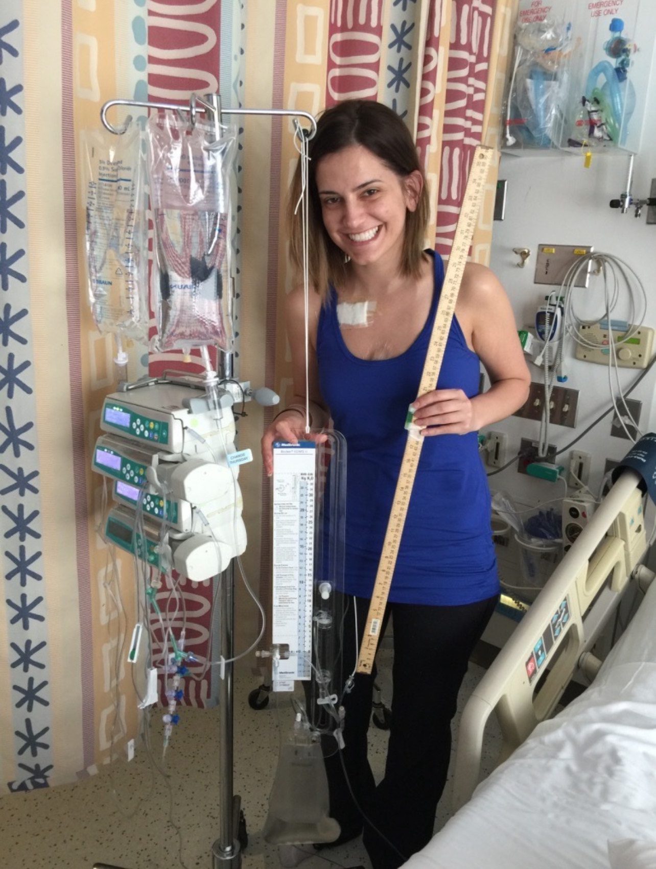
The EVD is connected to an external collection system that allows measurement and controlled drainage of the CSF.
Why and When are EVDs Used in Hydrocephalus?
EVDs are used in specific situations, including:
- Before Shunt Placement:
- When a patient suddenly develops hydrocephalus, an EVD can be placed immediately to help reduce pressure while doctors develop a plan for a more permanent treatment (such as placing a shunt, performing an endoscopic third ventriculostomy (ETV), or undertaking an operation to relieve the blockage – such as with tumors).
- During Shunt Malfunction (Shunt Failure):
- An EVD can be used as a temporary diversion of the CSF, to control the intracranial pressure while the shunt is being evaluated.
- Sometimes a shunt malfunction cannot be corrected immediately in the operating room, and placing an EVD provides the treating team time to come back and fix or replace the shunt.
- For Managing Infections:
- If a shunt becomes infected, it usually must be removed to effectively treat the infection; it can take many days to weeks for the infection to clear. If the patient still needs the CSF drained during that period, an EVD is placed to drain the CSF and allow completion of the infection treatment.
- Once the infection is gone, a new shunt is inserted.
- Assessing the need for a shunt (or determining the best pressure):
- In some cases, the EVD is used to determine the need for a shunt. The amount of drainage can be adjusted with the EVD, and the pressure can be directly measured, allowing the care team to see how a patient’s symptoms are affected.
- If as shunt is thought to be needed, sometimes the EVD can be adjusted in an attempt to define the best pressure setting for the shunt.
EVDs and Infections: Why and When They Are Used
Infections related to hydrocephalus, such as ventriculitis or infected shunts, require immediate treatment. An EVD plays an essential role in this process:
- Removing the Infected Shunt:
- It is usually necessary to completely remove the shunt hardware in order to make sure the infecting bacteria is removed.
- Placement of the EVD:
- Following shunt removal, an EVD is inserted to provide an external route for the CSF to drain, and to control the ICP.
- If there is infection in the CSF, placing an EVD and typically removing the shunt also prevents bacteria from continuing to contaminate the distal end of the shunt system, which could lead to infection in the the belly cavity (VP shunt) or the blood stream (VA shunt).
- Infection Management:
- The EVD allows doctors to regularly collect CSF samples to monitor how well the infection is clearing and to adjust treatment as necessary, ensuring the best possible recovery.
- Preparation for Shunt Replacement:
- After the infection has resolved, confirmed by several clear CSF tests, a new shunt can be placed to manage the hydrocephalus permanently.
By managing infections step by step, EVDs act as a temporary solution to diverting the CSF while the infection is treated, to test the CSF and the assess the effectiveness of the treatment, and to manage the pressure while the treatment is undertaken.
What to Expect While in the Hospital
If you or a loved one has an EVD, here is what you can expect:
- Placement:
- The EVD is inserted in a sterile environment, often in the operating room or intensive care unit.
- If a shunt is already present, the existing hole into the brain can often be used, but the old catheter is typically removed and a new catheter is used.
- Otherwise, a new small hole is drilled into the skull, and the tube is placed into the ventricle.
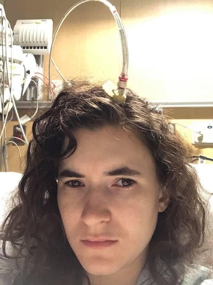
- The ventricular catheter is tunneled under the scalp and ‘externalized’ to be connected to the drain system. This tunneling helps to prevent infection from the scalp and to lessen the chance of the tubing being pulled out.
- Monitoring:
- Patients with an EVD are closely monitored to ensure it’s working properly.
- Medical staff also check the CSF for signs of infection.
- The drainage system can be adjusted (to control pressure) as needed.
- Duration:
- The length of time an EVD is in place depends on the individual’s condition. For an infection, it may be needed for several days to a couple of weeks. For bleeding, it may be used until the blood has cleared.
- CSF Sampling:
- Doctors may collect CSF samples from the EVD to check for infections and monitor treatment progress.
- Removal:
- Once the underlying problem has been corrected, then the EVD can be removed. This may be done at the same time the shunt is replaced. If a shunt is not needed, the EVD may be able to be removed at the bedside using aseptic techniques.
Risks and Complications
While EVDs are helpful and often life-saving, they come with risks, including:
- Infections:
- The tubing comes through the skin, so bacteria can track back into the CSF. This is why the system is kept ‘closed’, and the wounds are monitored along with sampling of the CSF.
- Over-Drainage or Under-Drainage:
- The amount of CSF drained is generally managed by the collecting device, by setting the collection tube at a certain height. When a patient moves relative to the system, or when they cough or talk, it can change that set pressure. This can result in too much or too little CSF being drained and, with that, symptoms can develop. The team will monitor not only the amount of CSF being drained, but also the patient’s condition.
- Blockages:
- The catheter (tube) can become clogged just like any shunt; this can require adjustment or replacement.
EVDs: A Critical Tool in Hydrocephalus Care
EVDs are one of many essential tools for managing hydrocephalus. They provide temporary relief from pressure in the brain and help doctors treat complications like infections or bleeding.
Knowing why EVDs are used, how they work, and what to expect during treatment can help patients and families feel more confident and prepared.


Information you can trust! This article was produced by the Hydrocephalus Association, copyright 2025. We would like to thank Bruce A. Kaufman, MD for his valuable contribution and expert input.