
Understanding Intracranial Pressure (ICP) Monitors in Hydrocephalus: When and Why They’re Used
For individuals with hydrocephalus, managing intracranial pressure (ICP) is a critical part of care.
ICP monitors are medical tools used to measure the pressure inside the skull, providing helpful information that assists doctors in making informed treatment decisions. This article explains why ICP monitors are used, how the procedure works, and their importance in managing hydrocephalus.
Why Are ICP Monitors Used in Hydrocephalus?

ICP monitors are used in specific situations to help doctors better understand and manage hydrocephalus. They may provide important data in the following scenarios:
- Diagnosing Symptoms: If someone is experiencing symptoms like severe headaches, nausea, or blurred vision, ICP monitoring can help identify if those are related to increased pressure. Sometimes very low pressure is the cause, and would not be found without measuring the pressure.
- Evaluating Shunt Function: Shunts can become blocked or malfunction or just not function well enough. ICP monitors provide direct pressure measurements that help define if the shunt is working as it should.
- Post-Surgery Monitoring: After surgery, ICP monitors can be used to follow the ICP, and ensure that the treatment is effective.
ICP monitors provide information on the pressure in a continuous way and in real time. This allows the doctors to identify trends in pressure before it causes a problem, or to correlate symptoms or findings with a specific pressure. The ultimate goal is to provide the correct treatment and to prevent injury to the brain due to pressure problems.
How Is ICP Monitoring Done?
The placement of an ICP monitor is a surgical procedure performed by a neurosurgeon. Here’s how the process works.
The type of ICP monitor determines how it is inserted:
- Subdural or Epidural Sensors: These monitors are placed on top of or just beneath the brain’s thick protective layer (the dura). They do not penetrate the brain but do provide pressure information – direct measurement if subdural (under the dura) or indirect if placed epidural (outside the dura, under the skull). They do not allow for the drainage of CSF.
- Intraventricular Catheter: A thin, flexible tube (often the same as a shunt catheter) is inserted into one of the brain’s fluid-filled spaces (ventricles). This method can also allow for draining excess cerebrospinal fluid (CSF).
- Fiberoptic Monitors: A thin (several millimeters – the size of a pencil lead) fiberoptic cable is inserted through the skull and dura, and into the brain tissue. This monitor can provide precise readings. These monitors do not allow for fluid drainage.
- Preparing for the Procedure
- Imaging Tests: Before surgery, imaging such as a CT or MRI scan may be used to determine the best location for placing the monitor.
- Anesthesia: The patient is sedated or given general anesthesia to ensure they are comfortable.
- Site preparation: The scalp is carefully cleaned to reduce the risk of infection.
- Creating the Hole in the skull: To place the monitor, the surgeon creates a small hole in the skull, called a burr hole or a twist drill hole.
- A small incision is made in the scalp to expose the skull.
- Using a surgical drill, the neurosurgeon creates a the hole in the skull. A ‘burr hole’ is about the size of a dime, while a ‘twist drill hole’ is about the size of small straw.
- If a monitor is being placed below the dura (subdural, intraventricular, or into the brain) then a small opening in the dura is created.
 Inserting the Monitor:
- The chosen monitor is then inserted to the desired location.
- Intraventricular catheters and subdural/epidural monitors are usually tunneled to leave the scalp a couple of centimeters from the insertion site (to reduce infection and chances of it coming out).
- Fiberoptic monitors are usually secured in place by a ‘bolt’ – a small metal device that is attached inside the hole in the skull and holds the monitor in place.
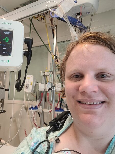
- Securing the Device and Continuous Monitoring: Once in place, the monitor is secured to prevent movement. It is connected to an external device that displays and can record pressure readings in real time. The incision is closed with stitches or staples.The monitor continuously tracks pressure levels, helping doctors make decisions about:
- Draining Excess CSF: If pressure is high, draining fluid is one way to treat the increased pressure.
- Additional Surgery: If abnormal pressure persists, the data may suggest the need for further intervention, like a shunt revision.
- Removing the Monitor: When monitoring is no longer required, the device is carefully removed:
- The monitor is withdrawn, and the incision site is cleaned and closed.
- The patient is monitored for signs of infection or discomfort as they recover.
Risks and Considerations
While ICP monitoring is generally safe, it does carry some risks, including:
- Infection: There is a small risk of infection where the monitor is placed.
- Bleeding: bleeding may occur during or after the procedure, and can be at the scalp, around the dura (outside or inside the dura), or even in the brain where the device is placed.
- Discomfort: Some patients may feel temporary soreness or irritation around the incision site.
Doctors take steps to minimize these risks and ensure close monitoring during and after the procedure.
Conclusion
ICP monitors are invaluable tools for managing hydrocephalus, providing real-time data to help doctors diagnose symptoms, evaluate shunt function, and plan treatment.
Understanding how these devices work can empower patients and families to take an active role in their care.
Information you can trust! This article was produced by the Hydrocephalus Association, copyright 2025. We would like to thank Bruce A. Kaufman, MD for his valuable contribution and expert input.